You are here
mri-ischemic-stroke.jpg

Media Folder
Title
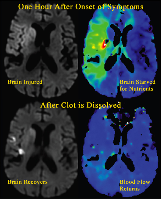
MRI images showing an ischemic stroke as it is happening and as it recovers
Caption
These MRI images show an ischemic stroke as it is happening and as it recovers. One hour after the onset of stroke symptoms, a region of brain is starved of blood because of clot in an artery and injured cells within the region light up on the stroke MRI. After the clot is dissolved by clot-busting drugs and blood returns to nourish the deprived brain region much of the injured brain recovers.
Credit
NINDS
Description
These MRI images show an ischemic stroke as it is happening and as it recovers. One hour after the onset of stroke symptoms, a region of brain is starved of blood because of clot in an artery [as seen in the colorized version of perfusion MRI] and injured cells within the region light up on the stroke MRI (as seen in the black and white diffusion weighted MRI). After the clot is dissolved by clot-busting drugs and blood returns to nourish the deprived brain region (second perfusion MRI) much of the injured brain recovers (second DWI).
Site_Section
Alt Text
MRI images showing an ischemic stroke as it is happening and as it recovers.
