You are here
20190122-nei-amd.jpg

Media Folder
Title
Images of mouse retinal scans
Caption
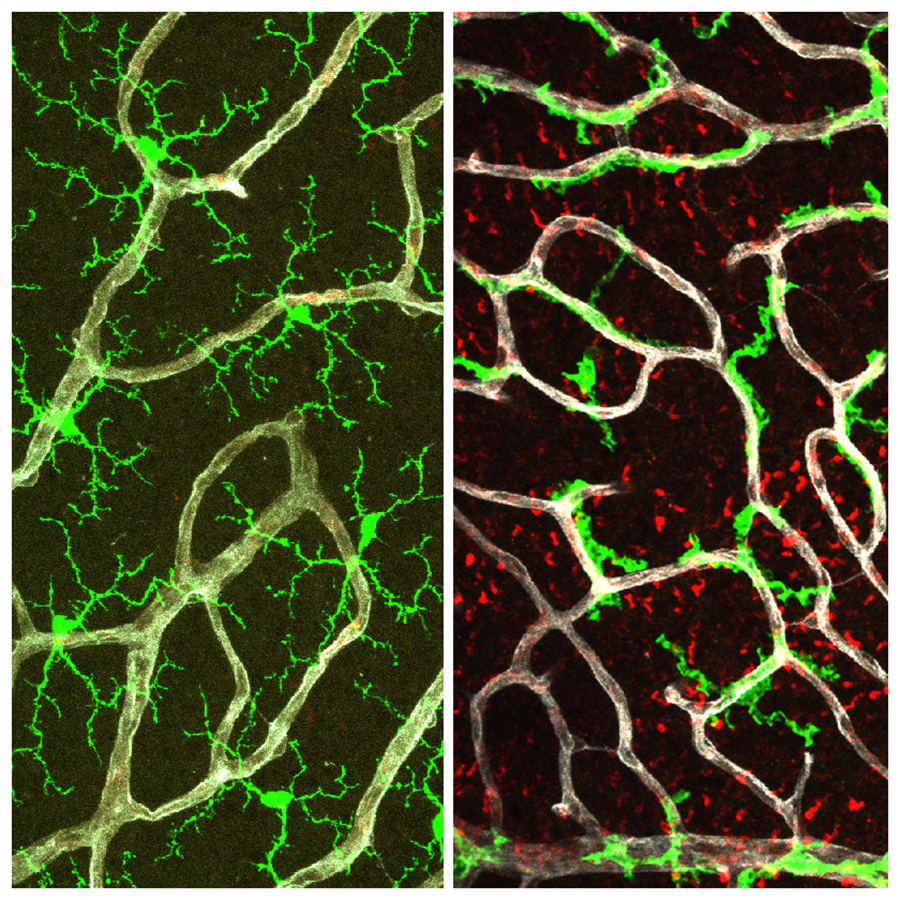
In healthy retina (left), microglia (green) demonstrate a branched structure that covers the retina. Without TGFβ signaling (right), microglia lose their branched structure, and attach to blood vessels (white). Müller glia become abnormal and acquire activation markers (red).
Credit
Wenxin Ma, M.D., Ph.D., and Wai Wong, M.D., Ph.D., National Eye Institute
Description
In healthy retina (left), microglia (green) demonstrate a branched structure that covers the retina. Without TGFβ signaling (right), microglia lose their branched structure, and attach to blood vessels (white). Müller glia become abnormal and acquire activation markers (red).
Site_Section
Alt Text
Images of mouse retinal scans.
