You are here
20190129-niaid-tick.jpg

Media Folder
Title
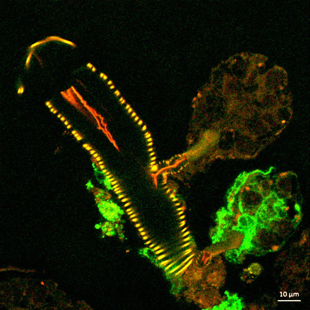
confocal microscope image shows a cross section of a tick salivary gland
Caption
This confocal microscope image shows a cross section of a tick salivary gland infected with Langat virus (green). Two rounded structures on the right, called acini, are shown attached to a duct (yellow). The lower acinus is infected, as denoted by the green fluorescent signal. (NIAID)
Credit
NIAID
Description
his confocal microscope image shows a cross section of a tick salivary gland infected with Langat virus (green). Two rounded structures on the right, called acini, are shown attached to a duct (yellow). The lower acinus is infected, as denoted by the green fluorescent signal.
Site_Section
Alt Text
Microscopic image of a tick salivary gland
