You are here
20210810-covid.jpg

Media Folder
Title
Colorized scanning electron micrograph of a cell (purple) heavily infected with SARS-COV-2 virus particles (yellow), isolated from a patient sample.
Caption
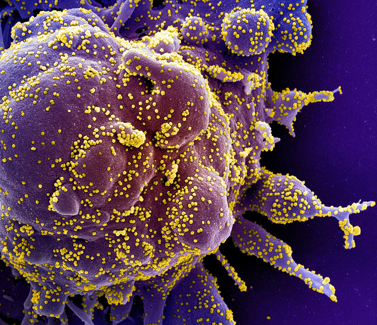
Colorized scanning electron micrograph of a cell (purple) heavily infected with SARS-COV-2 virus particles (yellow), isolated from a patient sample. Image captured at the NIAID Integrated Research Facility (IRF) in Fort Detrick, Maryland.
Credit
NIAID
Description
Colorized scanning electron micrograph of a cell (purple) heavily infected with SARS-COV-2 virus particles (yellow), isolated from a patient sample.
Site_Section
Alt Text
Colorized scanning electron micrograph of a cell (purple) heavily infected with SARS-COV-2 virus particles (yellow), isolated from a patient sample.
