You are here
20211202-eye.jpg

Media Folder
Title
Photographs of mouse retina showing the effect of uveitis treatment with i27-Bregs. The left column represents a normal retina. Photos in the middle and right column are retinal images from mice with uveitis, untreated or treated with i27-Bregs. The cent
Caption
Photographs of mouse retina showing the effect of uveitis treatment with i27-Bregs. The left column represents a normal retina. Photos in the middle and right column are retinal images from mice with uveitis, untreated or treated with i27-Bregs. The central spot is the optic nerve head. Note the absence of inflammation (ring surrounding the optic nerve) in the IL-27-treated retina (top right ima
Credit
Egwuagu Lab, NEI
Description
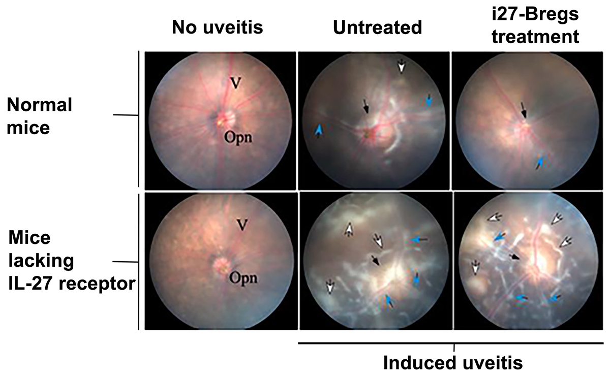
Photographs of mouse retina showing the effect of uveitis treatment with i27-Bregs. The left column represents a normal retina. Photos in the middle and right column are retinal images from mice with uveitis, untreated or treated with i27-Bregs. The central spot is the optic nerve head. Note the absence of inflammation (ring surrounding the optic nerve) in the IL-27-treated retina (top right image).
Site_Section
Alt Text
Photographs of mouse retina showing the effect of uveitis treatment with i27-Bregs. The left column represents a normal retina. Photos in the middle and right column are retinal images from mice with uveitis, untreated or treated with i27-Bregs. The cent
