You are here
20220308-lesion.jpg

Media Folder
Title
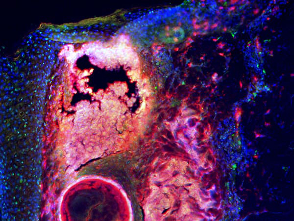
Inflamed area of pimple with central area appearing white
Caption
Microscopic image of an inflamed pimple with cathelicidin stained red, fat cells stained green and the nuclei of every cell stained blue. Because cathelicidin is produced from fat cells, their staining merges together.
Credit
Gallo lab, University of California, San Diego
Description
Inflamed area of pimple with central area appearing white
Site_Section
Alt Text
Inflamed area of pimple with central area appearing white
