You are here
PRinc_SB3769.jpg

Media Folder
Title
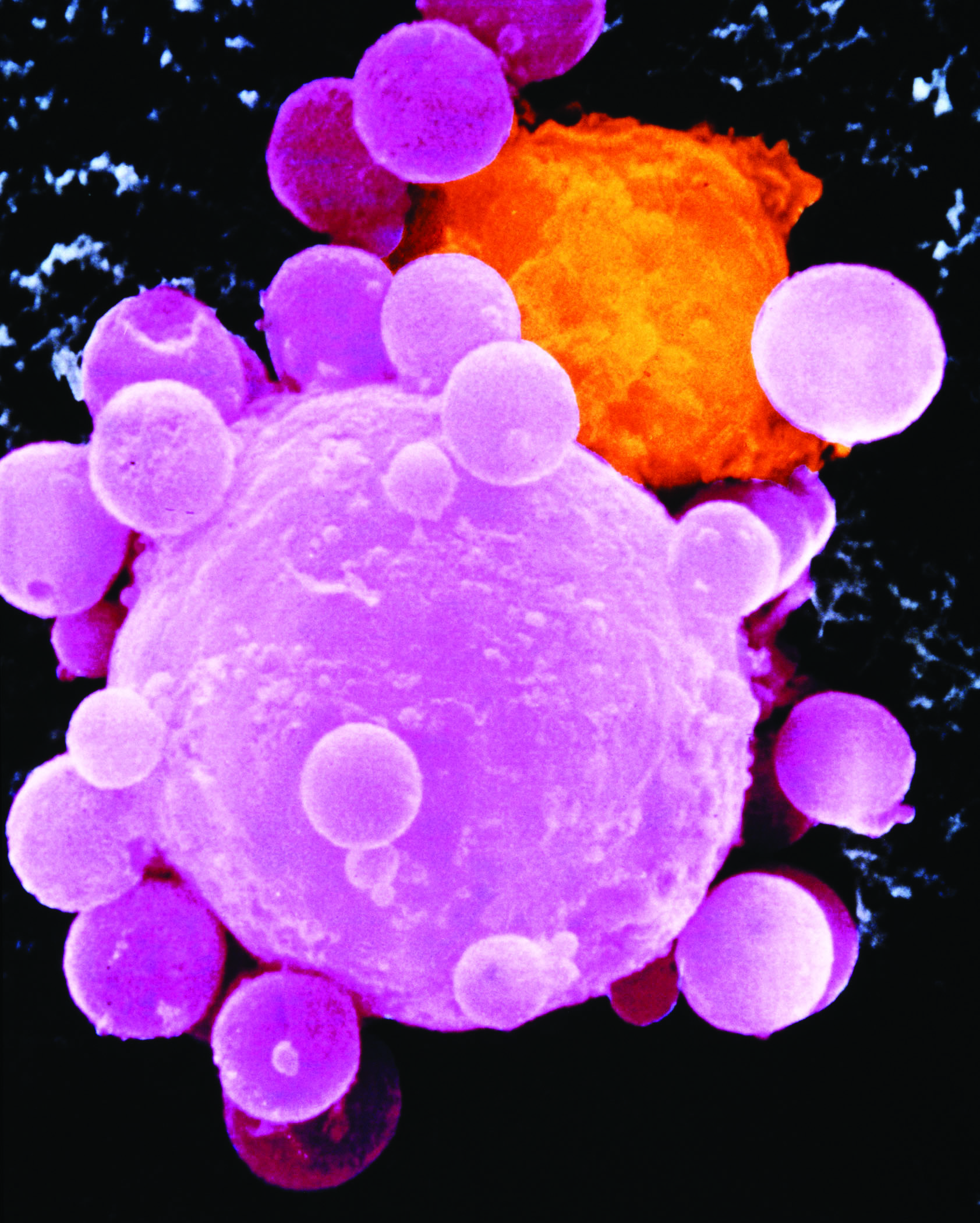
Colored scanning electron micrograph
Caption
Lung cancer cell division. Colored scanning electron micrograph (SEM) of a lung cancer cell during cell division (cytokinesis). The two daughter cells remain temporarily joined by a cytoplasmic bridge (centre). Cancer cells divide rapidly in a chaotic, uncontrolled manner. They may clump to form tumours, which invade and destroy surrounding tissues. Lung cancer is often associated with smoking tob
Credit
Steve Gschmeissner / Science Photo Library
Description
Lung cancer cell division. Colored scanning electron micrograph (SEM) of a lung cancer cell during cell division (cytokinesis). The two daughter cells remain temporarily joined by a cytoplasmic bridge (centre). Cancer cells divide rapidly in a chaotic, uncontrolled manner. They may clump to form tumours, which invade and destroy surrounding tissues. Lung cancer is often associated with smoking tobacco and exposure to industrial air pollutants. It causes a cough and chest pain and may spread to other areas of the body. Treatment includes removal of affected parts of the lung, with radiotherapy and chemotherapy. Magnification unknown.
Site_Section
Alt Text
Lung cancer cell division. Colored scanning electron micrograph of a lung cancer cell during cell division.
