
NIH scientists develop "digital twin" of eye cells to understand and treat age-related macular degeneration
Tuesday, February 10, 2026
NIH scientists develop "digital twin" of eye cells to understand and treat age-related macular degeneration
Breakthrough modeling technology reveals how cells lose their organization in leading cause of vision loss.
National Institutes of Health (NIH) researchers have developed a digital replica of crucial eye cells, providing a new tool for studying how the cells organize themselves when they are healthy and affected by diseases. The platform opens a new door for therapeutic discovery for blinding diseases such as age-related macular degeneration (AMD), a leading cause of vision loss in people over 50.
"This work represents the first ever subcellular resolution digital twin of a differentiated human primary cell, demonstrating how the eye is an ideal proving ground for developing methods that could be used more generally in biomedical research," Kapil Bharti, Ph.D., scientific director at the NIH’s National Eye Institute (NEI).
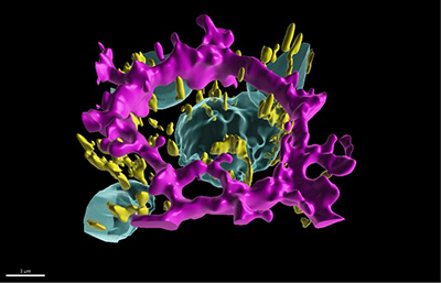
The researchers created a highly detailed, 3D data-driven digital twin of a retinal pigment epithelial (RPE) cells, which perform vital recycling and supportive roles to light-sensing photoreceptors in the retina. In diseases such as AMD, RPE cells die, which eventually leads to the death of photoreceptor cells, causing loss of vision.
For RPE cells to do their multiple jobs properly, they require a top-to-bottom polarity: The cell’s "top" (apical) side faces photoreceptors, where they recycle worn out photoreceptor parts daily. The cell’s "bottom"(basal) side faces the blood supply where it brings in nutrients and oxygen and ships out waste.
Researchers constructed the digital twin based on RPE cells made at NEI from induced pluripotent stem cells (iPS) cells developed by Allen Institute for Cell Science, Seattle. 3D imaging data for 1.3 million RPE cells, taken from nearly 4,000 fields of view cells, was collected using an automated confocal microscope.
Using the imaging data, the researchers trained an artificial intelligence (AI) algorithm they called polarity organization with learning-based analysis for RPE image segmentation, or POLARIS, to recognize the nucleus and other cell structures, and the cell’s shape and volume. 3D segmentation data (labels assigned to image voxels) were generated over different stages of cell development.
The researchers paid particular attention to polarity, quantifying the size and shape of the cell, its organelles and cytoskeletal structures including 3D spatial localization at various stages of development. They found that healthy, developing RPE cells follow a predictable path towards a polarized state.
The resulting AI-driven atlas of polarized and non-polarized RPE cells provides researchers with a reference for studying how diseases affect RPE at the cellular and subcellular levels, which could be transformative for therapeutic discovery.
“The digital twin approach represents a powerful new tool for AMD therapeutic development and could be adapted to study other eye and non-eye diseases and conditions affecting cell polarity,” said Bharti.
"By combining AI with mathematical modeling, we've created a window into cellular processes that were previously hidden from view," said the study’s first and a senior author, Davide Ortolan, Ph.D., NEI research follow. "This technology doesn't just help us understand what's happening in AMD, it gives us a platform to discover how to fix it."
This research was funded by the NIH/NEI Intramural Research Program.
NEI leads the federal government’s research on the visual system and eye diseases. NEI supports basic and clinical science programs to develop sight-saving treatments and address special needs of people with vision loss. For more information, visit https://www.nei.nih.gov.
About the National Institutes of Health (NIH): NIH, the nation's medical research agency, includes 27 Institutes and Centers and is a component of the U.S. Department of Health and Human Services. NIH is the primary federal agency conducting and supporting basic, clinical, and translational medical research, and is investigating the causes, treatments, and cures for both common and rare diseases. For more information about NIH and its programs, visit www.nih.gov.
NIH…Turning Discovery Into Health®
Reference
Ortolan D, Sathe P, Volkov A, Reichert D, Sebastian S, Maminishkis A, Schaub NJ, Ljungquist B, Bose D, Ferrari J, Lin N, Pegoraro G, Simon CG, Sharma R, Bajcsy P, and Bharti K. “AI driven 3D subcellular RPE map discovers cell state transitions in establishment of apical-basal polarity.” Published February 6, 2026 in Nature Partner Journal-AI. https://doi.org/10.1038/s44387-026-00074-6
