Scientists identify unique subtype of eczema linked to food allergy
Wednesday, February 20, 2019
Scientists identify unique subtype of eczema linked to food allergy

Atopic dermatitis, a common inflammatory skin condition also known as allergic eczema, affects nearly 20 percent of children, 30 percent of whom also have food allergies. Scientists have now found that children with both atopic dermatitis and food allergy have structural and molecular differences in the top layers of healthy-looking skin near the eczema lesions, whereas children with atopic dermatitis alone do not. Defining these differences may help identify children at elevated risk for developing food allergies, according to research published online today in Science Translational Medicine. The research was supported by the National Institute of Allergy and Infectious Diseases (NIAID), part of the National Institutes of Health.
“Children and families affected by food allergies must constantly guard against an accidental exposure to foods that could cause life-threatening allergic reactions,” said NIAID Director Anthony S. Fauci, M.D. “Eczema is a risk factor for developing food allergies, and thus early intervention to protect the skin may be one key to preventing food allergy.”
Children with atopic dermatitis develop patches of dry, itchy, scaly skin caused by allergic inflammation. Atopic dermatitis symptoms range from minor itchiness to extreme discomfort that can disrupt a child’s sleep and can lead to recurrent infections in scratched, broken skin.

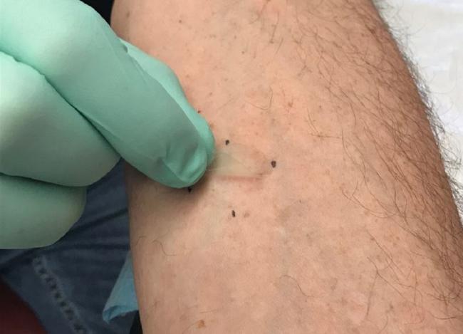
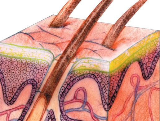
The study, led by Donald Y.M. Leung, M.D., Ph.D., of National Jewish Health in Denver, examined the top layers of the skin, known as the stratum corneum, in areas with eczema lesions and in adjacent normal-looking skin. The study enrolled 62 children aged 4 to 17 who either had atopic dermatitis and peanut allergy, atopic dermatitis and no evidence of any food allergy, or neither condition. Investigators collected skin samples by applying and removing small, sterile strips of tape to the same area of skin. With each removal, a microscopic sublayer of the first layer of skin tissue was collected and preserved for analysis. This technique allowed researchers to determine the skin’s composition of cells, proteins and fats, as well as its microbial communities, gene expression within skin cells and water loss through the skin barrier.
Researchers found that the skin rash of children with both atopic dermatitis and food allergy was indistinguishable from the skin rash of children with atopic dermatitis alone. However, they found significant differences in the structure and molecular composition of the top layer of non-lesional, healthy-appearing skin between children with atopic dermatitis and food allergy compared with children with atopic dermatitis alone. Non-lesional skin from children with atopic dermatitis and food allergy was more prone to water loss, had an abundance of the bacteria Staphylococcus aureus, and had gene expression typical of an immature skin barrier. These abnormalities also were seen in skin with active atopic dermatitis lesions, suggesting that skin abnormalities extend beyond the visible lesions in children with atopic dermatitis and food allergy but not in those with atopic dermatitis alone.
“Our team sought to understand how healthy-looking skin might be different in children who develop both atopic dermatitis and food allergy compared to children with atopic dermatitis alone,” said Dr. Leung. “Interestingly, we found those differences not within the skin rash but in samples of seemingly unaffected skin inches away. These insights may help us not only better understand atopic dermatitis, but also identify children most at risk for developing food allergies before they develop overt skin rash and, eventually, fine tune prevention strategies so fewer children are affected.”
Allergy experts consider atopic dermatitis to be an early step in the so-called “atopic march,” a common clinical progression found in some children in which atopic dermatitis progresses to food allergies and, sometimes, to respiratory allergies and allergic asthma. Many immunologists hypothesize that food allergens may reach immune cells more easily through a dysfunctional skin barrier affected by atopic dermatitis, thereby setting off biological processes that result in food allergies.
NIAID conducts and supports research—at NIH, throughout the United States, and worldwide—to study the causes of infectious and immune-mediated diseases, and to develop better means of preventing, diagnosing and treating these illnesses. News releases, fact sheets and other NIAID-related materials are available on the NIAID website.
About the National Institutes of Health (NIH): NIH, the nation's medical research agency, includes 27 Institutes and Centers and is a component of the U.S. Department of Health and Human Services. NIH is the primary federal agency conducting and supporting basic, clinical, and translational medical research, and is investigating the causes, treatments, and cures for both common and rare diseases. For more information about NIH and its programs, visit www.nih.gov.
NIH…Turning Discovery Into Health®
D Leung, et al. Non-lesional skin surface distinguishes atopic dermatitis with food allergy as unique endotype. Science Translational Medicine DOI: 10.1126/scitranslmed.aav2685 2019.
Institute/Center
Contact
301-402-1663


