
The BRAIN Initiative Photo and Video Contest
Brain Research Through Advancing Innovative Neurotechnologies® Initiative
The BRAIN Initiative Photo and Video Contest
The 2026 BRAIN Initiative Conference is hosting a photo and video contest. This unique contest is an opportunity to showcase the stunning, colorful, and inspiring images captured by today's advances in neurotechnology. We're excited to showcase the BRAIN community’s creative fusions of art and science!
Visit the 2026 BRAIN Initiative Conference website for information. Following the conference, this webpage will feature the 2026 winning photos and videos.
Past Winners
Explore the winners of previous year's entries.
2024
-
First place
Exploring the 3D nanoarchitecture of neuronal membranes at dendritic spine by labeling Channelrhodopsin-2
Image of Immunofluorescence-labeled Channelrhodopsin-2 (ChR2) in 50-μm thick tissue-clearing mouse brain slices captured using 4Pi single-molecule localization microscopy. The ChR2-decorated neuromembrane at the dendritic spine is outlined with sub-15-nm 3D resolution pseudocolored by depth position of proteins, revealing nanoscale view in the 3D architecture of the dendritic spine and its cross-sectional details.By Hao-Cheng Gao, Xi Cheng, Alexander Chubykin, and Fang Huang, Purdue University
-
Second place
Spinal motor neurons illuminated by enhancer AAV
Overview of a mouse spinal cord showing infectivity of an enhancer AAV driving SYFP2 expression (green) in spinal motor neurons following intravenous administration. The overview captures consistent expression throughout all levels of cord (cervical, thoracic, lumbar, and sacral) on both right and left hemispheres. The subsequent “fly-through” portion of the video stiches together images of ~2um thick transverse sections of the spinal cord progressing from top to bottom (cervical to sacral). SYFP2 expression matches the expected morphology and abundance of spinal motor neurons in the mouse spinal cord based off previous data, notably the Allen in situ hybridization (ISH) atlas.By Tanya Daigle and Emily Kussick, Allen Institute for Brain Science
-
Third place
Neuronal 'fireworks' when the salamander sniffs
In vivo real-time volumetric calcium imaging of salamander pallial neuron activity in response to odor stimuli with swept confocally-aligned planar excitation (SCAPE) microscopy.By Lu Xu, Wenze Li, Eliza Jaeger, Elizabeth Hillman, and Maria Tosches, Columbia University
-
BRAIN at 10 Video Finalist
Cortical Density
This animation reveals the immense scientific progress that has been made in visualizing the brain. The Golgi method, established around the year 1873, stains ~1-5% of neurons, leading to a view of neurons surrounded by much empty space. Modern methods detail the true complexity and density of brain tissue.By Tyler Sloan, Quorumetrix Studio
2023
-
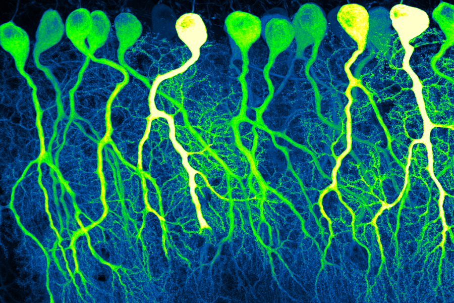
First Place Photo
Dark Commute at 4am
In the darkness of a confocal microscope room, bright fluorescent dyes reveal Purkinje cells winding their way through the tissue of the cerebellum. These complex, branching cells play roles in learning and memory. The cells in this photo, taken from sections of mouse cerebellum, resemble pre-dawn commuters on the highways of the brain as they travel towards their eventual targets.
By Silas Busch, University of Chicago
-
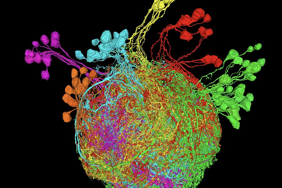
Second Place Photo
Premotor Neurons Controlling the Fruit Fly Leg
A reconstruction of premotor neurons controlling the fruit fly leg. Using an electron microscopy dataset of ultrathin sections of the Drosophila ventral nerve cord, researchers created a vivid display of the neural connections involved in fly leg movement. The structure of each neuron helps researchers determine their developmental lineages, represented by the different colors.
By Andrew Cook, Jasper Phelps, Anthony Azevedo, Ellen Lesser, Leila Elabbady, Brandon Pratt, Wei-Chung Allen Lee, John Tuthill, University of Washington and Harvard Medical School
-
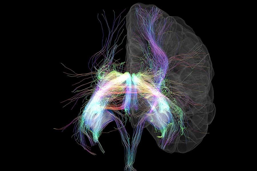
Third Place Photo
Memory Lanes
The hippocampus is the brain’s memory center. By combining two MRI scans, researchers can reveal the vast network of nerve fibers to and from the hippocampus—a wiring diagram for part of the brain. The axon fiber bundles are artificially colored depending on which direction they are heading. For a better sense of just how immensely complex the brain’s wiring is, this image represents less than 1% of the data collected.
By Tyler Ard, USC Stevens Neuroimaging and Informatics Institute


2022
-
First Place Video
Neurons In Action
Functional activity measured in vivo with 2-Photon imaging with matching morphologies from the same neurons measured with electron microscopy.By Andreas Tolias, Jacob Reimer, R.J. Cotton, Xaq Pitkow, Nuno da Costa, Forrest Collman, Clay Reid, and Sebastian Seung, Baylor College of Medicine, Allen Institute, Princeton University, Northwestern University/Shirley Ryan Ability Lab
2021
-
First Place Video
DBS Lead Placement for OCD
360 degree view of deep brain stimulation (DBS) lead placement in one participant that underwent DBS surgery for obsessive compulsive disorder (OCD).By Nicole Provenza, Raissa Mathura, Noam Peled, Evan Dastin-van Rijn, Kelly Bijanki, Sameer Sheth, David Borton, Wayne Goodman, Brown University, Baylor College of Medicine
-
Second Place Video (TIE)
Pyramidal Tract Reconstruction in Vivo
Pyramidal tract of an HCP subject reconstructed in vivo using Radial DSI and ODF-Fingerprinting. Improved reconstruction of fibers crossing at shallow angles ensured by ODF-Fingerprinting allowed to reproduce the reach fanning shape of cortical terminations of the tract. Images were rendered in DSI Studio.By Patryk Filipiak, Timothy Shepherd, Ying-Chia Lin, Dimitris G. Placantonakis, Fernando E. Boada, Steven H. Baete, New York University School of Medicine
-
Second Place Video (TIE)
Non-invasive in vivo Mapping of the Human Amygdala Circuit
Tractography of three critical amygdala pathways: the ventral amygdalofugal pathway, the stria terminalis and then amygdala-prefrontal pathway.By Josue Avecillas-Chasin, Ausaf Bari, Jean-Philippe Langevin, University of California, Los Angeles
This page last reviewed on
