
Protein may be linked to exercise intolerance in ME/CFS
September 12, 2023
Protein may be linked to exercise intolerance in ME/CFS
At a Glance
- A study suggested that high levels of a protein may reduce energy production in the muscle cells of people with ME/CFS.
- Blocking this protein in cells in the lab restored energy production, suggesting a potential new strategy for treating the condition.
Myalgic encephalomyelitis/chronic fatigue syndrome (ME/CFS) afflicts more than 2 million people nationwide. People with ME/CFS live with debilitating symptoms including exhaustion, exercise intolerance, cognitive problems, and a worsening of symptoms after even mild exertion (known as post-exertional malaise).
The causes of ME/CFS remain poorly understood, although many people first develop symptoms after a viral infection. This gap in understanding limits both diagnosis and the development of treatments.
A team of NIH researchers led by Drs. Paul Hwang, Avindra Nath, and Brian Walitt have been studying a woman who took days to recover after physical exertion and several of her relatives at the NIH Clinical Center. Their findings were published on August 22, 2023, in the Proceedings of the National Academy of Sciences.
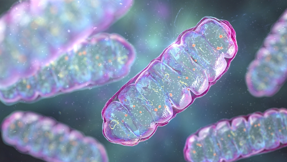
Tests done while the woman was exercising found a very slow recovery of cellular energy production after exertion. Muscle cells taken from the patient and examined in the lab showed reduced oxygen use. Oxygen is used by mitochondria, the cell compartment that makes energy molecules.
Further laboratory studies led the team to a protein called WASF3. This protein, which was boosted in response to cellular stress, disrupted the cells’ energy production. Blocking WASF3 allowed mitochondria to produce energy at normal levels. The team then showed that extra WASF3 in the cells interfered with formation of the structures that mitochondria use to produce energy.
To better understand the role of WASF3, the team engineered mice to produce excess WASF3. They found that, similar to people with post-exertional malaise, muscles in these mice were slow to recover after exercise. The mice also showed a 50% reduction in their ability to run on a treadmill, even though their muscle strength was comparable to mice without extra WASF3.
To see if WASF3 dysfunction might be involved in ME/CFS, the team compared muscle tissue samples taken from 14 people with ME/CFS to samples from 10 healthy volunteers. They found substantially higher levels of WASF3 in most of the people with ME/CFS.
This dysfunctional increase in WASF3 seemed to be linked to impairment of a cellular signaling pathway called the ER stress pathway. When the team treated human muscle cells with a compound known to increase ER stress, they saw a corresponding harmful increase in WASF3.
The researchers treated cells from the initial study participant with an experimental drug, called salubrinal, known to reduce ER stress. After this treatment, WASF3 levels decreased in the cells, more mitochondrial energy complexes formed, and energy production improved.
“We hope to embark on clinical studies to investigate whether this type of strategy can also work in patients to improve energy levels,” Hwang says.
Mitochondrial dysfunction has been found in some people with Long COVID and other conditions that include fatigue. More research is needed to understand whether targeting ER stress may also be a promising approach for these conditions.
—by Sharon Reynolds
Related Links
- Immune Cell Metabolism Altered in ME/CFS
- Blood Test May Detect Myalgic Encephalomyelitis/Chronic Fatigue Syndrome
- Feeling Fatigued? Finding Possible Causes
- Advancing ME/CFS Research
- Myalgic Encephalomyelitis/Chronic Fatigue Syndrome (CDC)
References
WASF3 disrupts mitochondrial respiration and may mediate exercise intolerance in myalgic encephalomyelitis/chronic fatigue syndrome. Wang PY, Ma J, Kim YC, Son AY, Syed AM, Liu C, Mori MP, Huffstutler RD, Stolinski JL, Talagala SL, Kang JG, Walitt BT, Nath A, Hwang PM. Proc Natl Acad Sci U S A. 2023 Aug 22;120(34):e2302738120. doi: 10.1073/pnas.2302738120. Epub 2023 Aug 14. PMID: 37579159.
Funding
NIH’s National Heart, Lung, and Blood Institute (NHLBI) and National Institute of Neurological Disorders and Stroke (NINDS).
