Tissue regeneration to help healing
Research in Context November 17, 2026
Tissue regeneration to help healing
Replacing damaged cells to treat injury and disease
To stay alive, we have to replace cells that aren’t doing their job well, a process called regeneration. This Research in Context feature examines some of the ways NIH-supported scientists are trying to help our bodies do that through regenerative medicine.
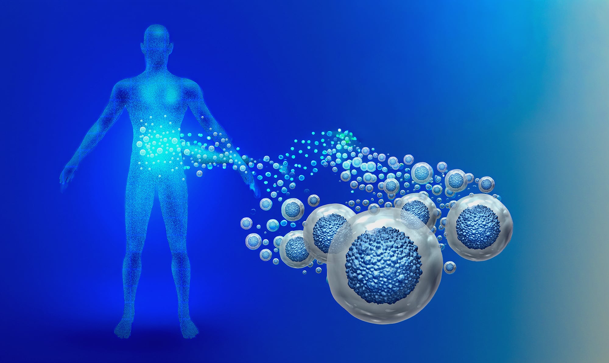
In healthy people, the body runs like a well-oiled machine. Specialized tissues and organs do important jobs like digesting food and removing toxins from the blood. Aging, injury, and disease can all damage tissue and make it worse at doing its job.
To fix that, scientists are exploring ways to replace tissue that isn’t working well with new, healthy tissue. Some studies look at natural healing to figure out how to boost regeneration without scarring. Others look at stem cells that serve as the seeds for new tissue to grow. Scientists are also trying to grow healthy tissue in the lab to be transplanted back into the body.
But no matter where the tissue comes from, this area of research—known as tissue regeneration—could one day remake the way we recover from injury and disease.
How healing happens
The immune system is critical for mending tissue damage. One way this system helps tissue repair is by causing inflammation. Unchecked, inflammation can be harmful. But normal, healthy inflammation promotes tissue healing.
“It’s a Goldilocks model,” explains Dr. Shruti Naik of the Icahn School of Medicine at Mount Sinai. “You don’t want too little because then you’re not going to kickstart healing, but you also don’t want so much that your body thinks there’s a five-alarm fire.”
Inflammation recruits immune cells that fight infection and get rid of dead or damaged cells. Immune cells also prompt the remaining cells in damaged tissue to make more of themselves.
“Immune cells help the other cells of the body adopt features needed for healing,” Naik says.
One of those features is boosting energy. Naik’s NIH-funded research team has found that immune cells in damaged skin release a molecule called interleukin-17A. It promotes relocation of nearby skin cells to the damaged tissue to help fuel tissue repair. Studies like this point to treatment targets that might help tissues recover from damage.
The body’s supply lines
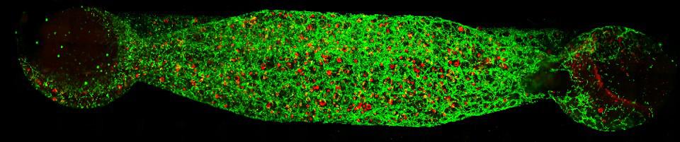
Like any living thing, tissue needs to be nourished. So new tissue requires new blood vessels. Blood flows through these vessels to bring oxygen and nutrients to tissue.
Immune cells encourage new blood vessels to grow by releasing a molecule called vascular endothelial growth factor A. This molecule spurs the cells that line the inside of blood vessels, called endothelial cells, to make more of themselves.
But these endothelial cells “aren’t just passive plumbing,” for our blood, says Dr. Shahin Rafii of Weill Cornell Medicine. They also release unique and organ-specific growth factors that help with regeneration. Rafii’s NIH-funded research has pioneered the study of those chemicals.
“Tissues without working and healthy blood vessels do not exist and will scar,” he notes.
A better understanding of the endothelial cells that make up blood vessels could provide critical insights into how tissues heal. It could also help in developing strategies to promote organ repair without scarring.
Stopping scars
Regeneration isn’t just about growing and feeding the new tissue we need. It’s also about stopping the growth of tissue we don’t want.
Scar tissue can grow in any part of the body where damage has occurred. Scars form in a process called fibrosis. It’s caused by cells called fibroblasts laying down too much of a network of molecules outside of cells called extracellular matrix.
“The fundamental purpose of scar tissue is to close wounds fast so you don’t die,” explains Dr. Kaitlyn Sadtler of NIH’s National Institute of Biomedical Imaging and Bioengineering (NIBIB). “You don’t want your bowel leaking bacteria into your gut if there’s damage there, and you don’t want your internal organs to be bleeding a lot, so fibrosis makes sense. It might be like plopping a bandage over a bullet hole, but at least the wound is closed.”
Scar tissue provides important structural support to damaged tissue, but it can’t do that tissue’s regular job. It’s also a lot stiffer. As a result, runaway fibrosis prevents organs from working normally.
Although most people don’t know the word fibrosis, it contributes to almost 45% of all deaths in the U.S., according to Dr. Michael Longaker, an expert on fibrosis at Stanford University.
Inflammation influences how much fibrosis happens after tissue damage. But the mechanical pushing and pulling forces that occur in damaged tissue are also involved.
“The crosstalk between mechanical forces and the immune system revs up fibroblasts,” Longaker explains. “If we interrupt that, we might see less scarring.”
In a recent NIH-funded study, Longaker’s team and collaborators identified specific fibroblasts that are activated by mechanical forces in the skin. They were able to reduce skin scarring by suppressing the process of fibrosis.
Fibrosis in the skin and within the body happens in a similar way. “There are not a million ways to form a scar,” Longaker notes. So the team’s discoveries might also help reduce fibrosis when internal organs suffer damage.
Sadtler’s lab at NIH is seeking to curb fibrosis using cutting-edge materials that surgeons use to repair tissue. By learning why certain materials encourage or prevent fibrosis during healing, her team hopes to create therapies that mimic how the beneficial materials affect regeneration.
In a study published in 2024, Sadtler’s team found that muscle injuries in mice healed better with less fibrosis when treated with extracellular matrix material from which cells have been removed. The body gradually replaced the material with healthy, functional tissue.
The matrix also lured more immune cells called cDC1 cells to the damaged muscle, where they calmed inflammation. When mice didn’t have cDC1 cells, they replaced more of the injured muscle with scar tissue, bone, and fat.
“cDC1 cells are really important for regulating that immune response early on after traumatic injury,” Sadtler says.
Discoveries like those made in Longaker’s and Sadtler’s labs could one day lead to treatments that reduce or prevent the formation of scar tissue as injuries heal.
Studying stem cells
All the cells that make up an embryo growing in the womb come from an initial group of cells called stem cells. Stem cells can both make copies of themselves and transform into other types of cells.
An embryo’s stem cells give it amazing regenerative abilities. Longaker’s research has revealed that if an embryo is damaged within about six months of conception, it will heal without scarring.
Later in life, the body uses stem cells to replace damaged tissue. To help that process along, scientists need to determine which stem cells the body uses to regrow specific tissues.
NIH-funded scientists have identified several sets of stem cells that help different tissues heal, including stem cells that repair damage in the mouth and the lungs. Longaker and his Stanford University colleague Dr. Charles Chan identified the specific stem cells that generate new bone. Those cells also produce supportive tissue called stroma and the cartilage that cushions our joints.
“The ultimate blueprint for organ and tissue regeneration is how they develop in utero, so if you understand the stem cells that are involved in how that tissue first forms, I think it gives you the best shot to recreate the response to injury during development and get the injury to regenerate and not scar,” Longaker says.
Growing tissue outside the body
Sometimes, there aren’t enough stem cells in the body to repair damaged tissue, so many scientists are trying to grow healthy tissue in the lab. This could then replace ailing tissue in the body.
One recent NIH-funded study tested a way to treat liver damage with lab-grown tissue. Led by Drs. Sangeeta Bhatia of the Massachusetts Institute of Technology and Christopher Chen of Boston University and the Wyss Institute, the researchers grew a small batch of liver tissue in the lab that was genetically altered so they could control its growth. After the liver tissue was placed into mice, it only grew when the mice were given a specific compound.
Since the researchers could make the liver tissue grow after placing it into the body, treating liver damage in this way would only require growing a small amount of tissue in the lab. That’s much easier than growing a large piece of tissue and keeping it alive outside the body.
After tissue moves from lab to patient, endothelial cells may help it function better, according to Rafii. His lab is currently using endothelial cells in an experimental treatment for type 1 diabetes. This disease occurs because patients have too little of the hormone insulin, which helps control blood sugar. Cells called islets in the pancreas make insulin.
In an NIH-funded study published in 2025, Rafii’s team transplanted islets from human donors and engineered human endothelial cells into mouse models of type 1 diabetes. The treatment successfully brought down the mice’s blood sugar levels.
“Without these customized endothelial cells, the islets don’t function,” Rafii says. “If I just transplant the islets, they don’t last long. If I transplant them with the these programmed endothelial cells, they engraft more durably and regulate blood sugar more effectively.”
Eventually, Rafii wants to figure out a way to take stem cells from a patient’s intestines or human pluripotent stem cells, turn them into islets, and transplant them back into the patient’s body. A patient’s immune system is less likely to attack cells grown using their own stem cells than stem cells from another person.
But keeping stem cells alive outside the body is difficult. Rafii believes pairing stem cells with customized endothelial cells might help there as well.
“These organ-specific endothelial cells provide the right signals to the stem cells,” he says. “It’s like a father and mother nurturing a young child.”
The long road to lab-grown organism
As difficult as it is to grow healthy tissue outside the body, it’s even harder to grow entire organs. But with nearly 100,000 Americans waiting to receive an organ transplant, researchers have been trying to overcome that challenge.
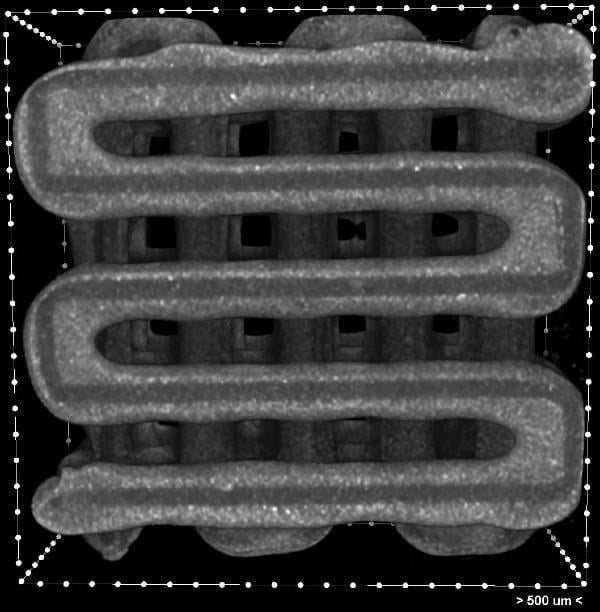
Scientists are moving closer to growing organs in the lab by creating structures called scaffolds. Like the temporary scaffolds that surround buildings under construction, biological scaffolds support lab-grown organs as they form. Some of them incorporate molecules that help cells grow. If the scaffold is biodegradable, the body can break it down after the organ is transplanted into a patient, leaving just the new organ. For this reason, many advanced scaffolds are made of the same extracellular matrix components that Sadtler’s lab is studying.
One of the barriers to growing organs in the lab is the fact that organs are made of multiple types of cells, and it’s hard to grow a variety of cells on a single scaffold. A research team led by Dr. Antonios Mikos of Rice University created a technique that could make it easier to do that. By carving grooves into the fibers that make up scaffolds, the scientists can build scaffolds with multiple layers. Each layer can then support the growth of a different type of cell.
Researchers are also growing miniature organs called organoids in the lab as a stepping stone towards creating full-size ones. Organoids are tiny three-dimensional collections of the types of cells found in a particular organ. They even behave similarly to real organs.
While organoids are not perfect replicas of real organs, they’re much more similar to human physiology than tissue from mice, Rafii says. That makes them a great tool for learning about how organs grow and repair themselves.
And just like stem cells and islet cells, organoids benefit from being grown alongside endothelial cells. “We have to give them blood vessels to get them to act like actual human tissue,” Rafii says.
“The more we understand the conversation between these specialized endothelial cells and the tissue-specific organoids, the more we can understand how the body regenerates itself without complications associated with fibrosis,” he adds.
Future directions
Lab-grown replacement organs and shots that instantly heal injuries are still a long way off. But NIH-funded research is bringing them closer to reality. Researchers are learning how the body regrows tissue and exploring ways to grow new tissue outside the body.
At the same time, the field is expanding beyond our own cells. Scientists have begun to consider the role of the trillions of microbes that live on and inside our body as well.
“Microbes can help educate the immune system in a positive way to promote healing,” Naik says.
As research progresses, scientists hope to figure out ways to help people recover from injury and disease faster with less scarring and disability.
“I think it’s a really exciting time in regenerative medicine because the technologies are finally online that will let us push the needle and heal the body,” Naik says. “Medicine has been focused on halting disease, and regenerative medicine is going to bring people back to good health.”
—by Brandon Levy
Related Links
- Scientists spur growth of implanted liver tissue
- Scientists create organoids with specialized blood vessels
- Bioengineered vessels transform into living blood vessels
- Newly identified oral stem cell key to wound healing
- Human skeletal stem cell identified
- Tendon trouble
- To heal a wound
- Regenerative medicine innovation project
- Regeneration
- NIH stem cell information
- Organ donation statistics (HRSA)
References
Targeting circulating mechanoresponsive monocytes and macrophages to reduce fibrosis. Chen K, Griffin M, Henn D, Berryman KS, Sivaraj D, Kussie HC, Bonham CA, McKenna E, Mora Pinos MG, Alsharif AM, Saenz F, Matthews NE, Jafri M, Yasmeh JP, Borrelli MR, Huskins S, Steele S, Knochel AB, Singh M, Hostler AC, Leeolou M, Hahn WW, Perrault D, Abbas D, Guardino N, Thomas B, Barrera JA, Noishiki C, Trotsyuk AA, Padmanabhan J, Januszyk M, Khreiss M, Longaker MT, Gurtner GC. Nat Biomed Eng. 2026 Jun;10(6):1247-1262. doi: 10.1038/s41551-025-01479-5. Epub 2025 Oct 21. PMID: 41120571.
Synthetic control of implanted engineered liver tissue growth. Stoddard AE, Kumar V, Tzouanas CN, Hui V, Li J, Jain A, Farrell A, Bhatia SN, Chen CS. Sci Adv. 2026 Apr 17;12(16):eadz8362. doi: 10.1126/sciadv.adz8362. Epub 2026 Apr 17. PMID: 41996502.
Vascularization of human islets by adaptable endothelium for durable and functional subcutaneous engraftment. Li G, Craig-Schapiro R, Redmond D, Chen K, Lin Y, Geng F, Gao M, Rabbany SY, Suresh G, Pearson B, Schreiner R, Rafii S. Sci Adv. 2025 Jan 31;11(5):eadq5302. doi: 10.1126/sciadv.adq5302. Epub 2025 Jan 29. PMID: 39879286.
Pro-regenerative biomaterials recruit immunoregulatory dendritic cells after traumatic injury. Lokwani R, Josyula A, Ngo TB, DeStefano S, Fertil D, Faust M, Adusei KM, Bhuiyan M, Lin A, Karkanitsa M, Maclean E, Fathi P, Su Y, Liu J, Vishwasrao HD, Sadtler K. Nat Mater. 2024 Jan;23(1):147-157. doi: 10.1038/s41563-023-01689-9. Epub 2023 Oct 23. PMID: 37872423.
Immune-Epithelial Cross Talk in Regeneration and Repair. Guenin-Mace L, Konieczny P, Naik S. Annu Rev Immunol. 2023 Apr 26;41:207-228. doi: 10.1146/annurev-immunol-101721-062818. Epub 2023 Jan 25. PMID: 36696569.
Emerging frontiers in regenerative medicine. McKinley KL, Longaker MT, Naik S. Science. 2023 May 26;380(6647):796-798. doi: 10.1126/science.add6492. Epub 2023 May 25. PMID: 37228215.
Interleukin-17 governs hypoxic adaptation of injured epithelium. Konieczny P, Xing Y, Sidhu I, Subudhi I, Mansfield KP, Hsieh B, Biancur DE, Larsen SB, Cammer M, Li D, Landén NX, Loomis C, Heguy A, Tikhonova AN, Tsirigos A, Naik S. Science. 2022 Jul 8;377(6602):eabg9302. doi: 10.1126/science.abg9302. PMID: 35709248.
Fiber engraving for bioink bioprinting within 3D printed tissue engineering scaffolds. Diaz-Gomez L, Elizondo ME, Koons GL, Diba M, Chim LK, Cosgriff-Hernandez E, Melchiorri AJ, Mikos AG. Bioprinting. 2020 Jun;18:e00076. doi: 10.1016/j.bprint.2020.e00076. Epub 2020 Jan 10. PMID: 33693067.
Heterogeneity within stratified epithelial stem cell populations maintains the oral mucosa in response to physiological stress. Byrd KM, Piehl NC, Patel JH, Huh WJ, Sequeira I, Lough KJ, Wagner BL, Marangoni P, Watt FM, Klein OD, Coffey RJ, Williams SE. Cell Stem Cell. 2019 Dec 5;25(6):814-829.e6. doi: 10.1016/j.stem.2019.11.005. PMID: 31809739.
Identification of the human skeletal stem cell. Chan CKF, Gulati GS, Sinha R, Tompkins JV, Lopez M, Carter AC, Ransom RC, Reinisch A, Wearda T, Murphy M, Brewer RE, Koepke LS, Marecic O, Manjunath A, Seo EY, Leavitt T, Lu WJ, Nguyen A, Conley SD, Salhotra A, Ambrosi TH, Borrelli MR, Siebel T, Chan K, Schallmoser K, Seita J, Sahoo D, Goodnough H, Bishop J, Gardner M, Majeti R, Wan DC, Goodman S, Weissman IL, Chang HY, Longaker MT. Cell. 2018 Sep 20;175(1):43-56.e21. doi: 10.1016/j.cell.2018.07.029. PMID: 30241615.
Regeneration of the lung alveolus by an evolutionarily conserved epithelial progenitor. Zacharias WJ, Frank DB, Zepp JA, Morley MP, Alkhaleel FA, Kong J, Zhou S, Cantu E, Morrisey EE. Nature. 2018 Feb 28. doi: 10.1038/nature25786. [Epub ahead of print]. PMID: 29489752.