You are here
20181204-prion.jpg

Media Folder
Title
Two panel image of retinas
Caption
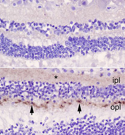
Top, retina of a control patient. Bottom, retina from a patient with CJD. Arrowheads point to abnormal prions in the outer plexiform layer (opl), and the asterisk (*) marks more diffuse prions in the inner plexiform layer (ipl).
Credit
Orrù et al., mBio
Description
Top, retina of a control patient. Bottom, retina from a patient with CJD. Arrowheads point to abnormal prions in the outer plexiform layer (opl), and the asterisk (*) marks more diffuse prions in the inner plexiform layer (ipl).
Site_Section
Alt Text
Two panel image of retinas
