You are here
20190604-brain.jpg

Media Folder
Title
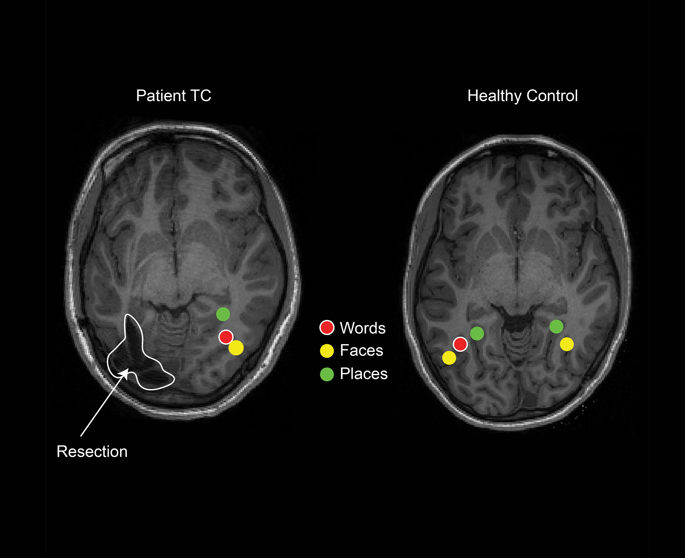
fMRI scans show that patient TC’s word-specific region, which is normally found on the left, has remapped to the right hemisphere.
Caption
fMRI scans show that patient TC’s word-specific region, which is normally found on the left, has remapped to the right hemisphere.
Credit
Erez Freud, Ph.D., York University.
Description
2D MRI brain scans of patient TC and a healthy control child reveal word, place, and face specific regions. Patient TC shows all three on the right, and resected region on the left. Healthy control has all three on the left, plus face and place on the right.
Site_Section
Alt Text
2D MRI brain scans of patient TC and a healthy control child reveal word, place, and face specific regions. Patient TC shows all three on the right, and resected region on the left. Healthy control has all three on the left, plus face and place on the rig
