You are here
20230314-emmi.png

Media Folder
Title
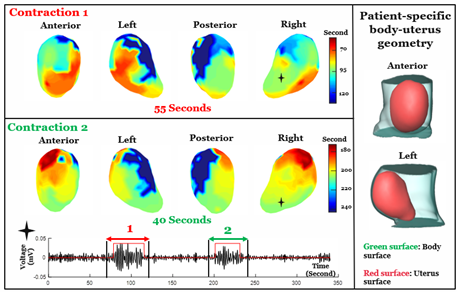
Scientific image measuring two contraction examples showing the voltage of the contraction and how long the contraction lasted, showing the output that can be generated using the EMMI technique.
Caption
: In this example, two adjacent contractions were imaged during labor. EMMI shows that the first contraction starts from the middle segment of the uterus and propagates up and down simultaneously. The second contraction starts from the top of uterus and moves in a faster and more synchronized manner than the first contraction.
Credit
Washington University in St. Louis
Description
Scientific image measuring two contraction examples showing the voltage of the contraction and how long the contraction lasted, showing the output that can be generated using the EMMI technique.
Site_Section
Alt Text
Scientific image measuring two contraction examples showing the voltage of the contraction and how long the contraction lasted, showing the output that can be generated using the EMMI technique.
