You are here
20231003-wounds.jpg

Media Folder
Title
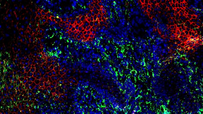
Immunofluorescence image with an area of disordered cells.
Caption
Immunofluorescence image of the edge of a diabetic wound. Two proteins involved in the release of exosomes from keratinocytes are shown in red and green.
Credit
Subhadip Ghatak
Description
Immunofluorescence image with an area of disordered cells.
Site_Section
Alt Text
Immunofluorescence image with an area of disordered cells.
