You are here
20250423-tam-ai.jpg

Media Folder
Title
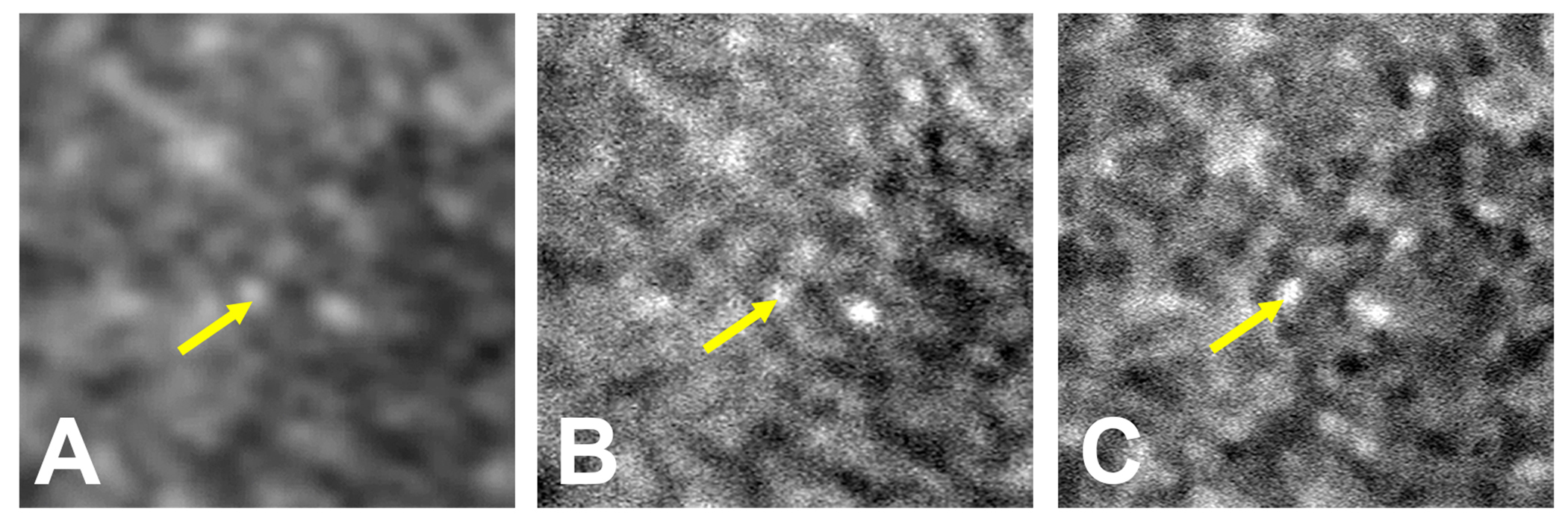
3 slides of retina tissue taken using three separate imaging techniques. Each image gets progressively clearer.
Caption
Comparison of the same patch of retina labeled with indocyanine green and visualized 3 different ways. A) Scanning laser ophthalmoscopy. B) AI-enhanced scanning laser ophthalmoscopy. C) Adaptive optics scanning laser ophthalmoscopy. Arrows highlight the same cell seen in different modalities.
Description
3 slides of retina tissue taken using three separate imaging techniques. Each image gets progressively clearer.
Site_Section
Alt Text
3 slides of retina tissue taken using three separate imaging techniques. Each image gets progressively clearer.
