NIH funds three national cryo-EM service centers and training for new microscopists
Tuesday, May 15, 2018
NIH funds three national cryo-EM service centers and training for new microscopists

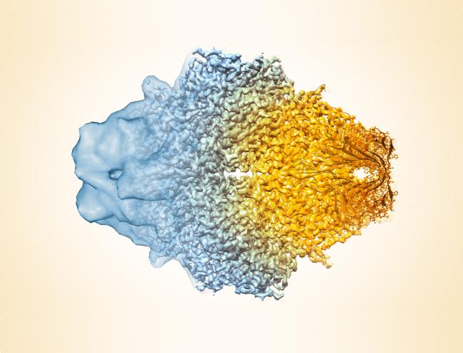
The National Institutes of Health is supporting efforts to broaden biomedical scientists’ access to cryo-electron microscopy (cryo-EM), the Nobel Prize-winning imaging method that is revolutionizing structural biology. The Transformative High Resolution Cryo-Electron Microscopy program is creating three national cryo-EM service centers to provide access to the technology and is supporting the development of cryo-EM training curricula to build a skilled workforce. The awards are anticipated to total $129.5 million, pending the availability of funds, and the centers are expected to offer limited services by late 2018 as they build to full capacity.
Microscopy is an important tool for scientists in the study of cells, tissues, and organs. Cryo-EM is a method used to image frozen biological molecules without the use of structure-altering dyes or fixatives or the need for crystallization to provide a more accurate model of the molecules and a greater understanding of biological function. Recent advances in cryo-EM technology have made it possible for scientists to obtain detailed images and structures of many biological molecules that cannot be obtained using other methods, like X-ray crystallography. Despite the emergence of cryo- EM as a powerful high-resolution imaging method, its use is hampered by inadequate access to equipment and a limited workforce.
“Cryo-electron microscopy is allowing us to resolve the three-dimensional structures of important biomolecules involved in disease that were inaccessible using previous technologies,” said National Institute of General Medical Sciences Director Jon R. Lorsch, Ph.D. “NIH wants to ensure as many scientists as possible have access to this crucial technology.”
The three national centers will be established with six-year awards at the New York Structural Biology Center, New York City; the Oregon Health & Science University, Portland, in partnership with the Pacific Northwest National Laboratory, Richland, Washington; and the SLAC National Accelerator Laboratory at Stanford University, Menlo Park, California. The centers will provide scientists with access to state-of-the-art cryo-EM technology and training, from sample preparation to collection of high-resolution data and computational analysis.
An important component of the program is to increase hands-on training and the availability of instructional material on cryo-EM methodology, which lags the development of the technology.
“The method is only as useful as the hands behind it. We need to ensure we have a well-trained, highly skilled workforce ready to meet the demands for cryo-EM,” said National Eye Institute Director Paul A. Sieving, M.D., Ph.D.
Four, three-year grants for the creation of instructional and hands-on training in cryo-EM methodology were awarded to the California Institute of Technology, Pasadena, California; Yale University, New Haven, Connecticut; the University of Utah, Salt Lake City; and Purdue University, West Lafayette, Indiana. The grants will be used to develop online video lectures, software and e-books, 3-D animations and interactive simulations, and interactive virtual reality to train novice and experienced users on cryo-EM technology and theory.
The Transformative High Resolution Cryo-Electron Microscopy program is funded by the NIH Common Fund, managed by a trans-NIH working group, and led by staff from the Common Fund, the National Institute of General Medical Sciences, and the National Eye Institute.
“The NIH Common Fund focuses on speeding up discovery in the biomedical sciences,” said James M. Anderson, M.D., Ph.D., director of the Division of Program Coordination, Planning, and Strategic Initiatives, which oversees the NIH Common Fund. “By increasing access to cryo-EM technology and training, we hope to spur discoveries across the spectrum of biomedical science from basic to clinical research, and in multiple disease areas.”
About the NIH Common Fund: The NIH Common Fund encourages collaboration and supports a series of exceptionally high-impact, trans-NIH programs. Common Fund programs are managed by the Office of Strategic Coordination in the Division of Program Coordination, Planning, and Strategic Initiatives in the NIH Office of the Director in partnership with the NIH Institutes, Centers, and Offices. More information is available at the Common Fund website: https://commonfund.nih.gov.
About the National Institute of General Medical Sciences (NIGMS): NIGMS supports basic research that increases our understanding of biological processes and lays the foundation for advances in disease diagnosis, treatment, and prevention. NIGMS-funded scientists investigate how living systems work at a range of levels from molecules and cells to tissues and organs, in research organisms, humans, and populations. Additionally, to ensure the vitality and continued productivity of the research enterprise, NIGMS provides leadership in training the next generation of scientists, in enhancing the diversity of the scientific workforce, and in developing research capacity throughout the country.
About the National Eye Institute (NEI): NEI leads the federal government’s research on the visual system and eye diseases. NEI supports basic and clinical science programs to develop sight-saving treatments and address special needs of people with vision loss. For more information, visit https://www.nei.nih.gov.
About the National Institutes of Health (NIH): NIH, the nation's medical research agency, includes 27 Institutes and Centers and is a component of the U.S. Department of Health and Human Services. NIH is the primary federal agency conducting and supporting basic, clinical, and translational medical research, and is investigating the causes, treatments, and cures for both common and rare diseases. For more information about NIH and its programs, visit www.nih.gov.
NIH…Turning Discovery Into Health®
Institute/Center
Contact
301-435-0968


